Ga offline met de app Player FM !
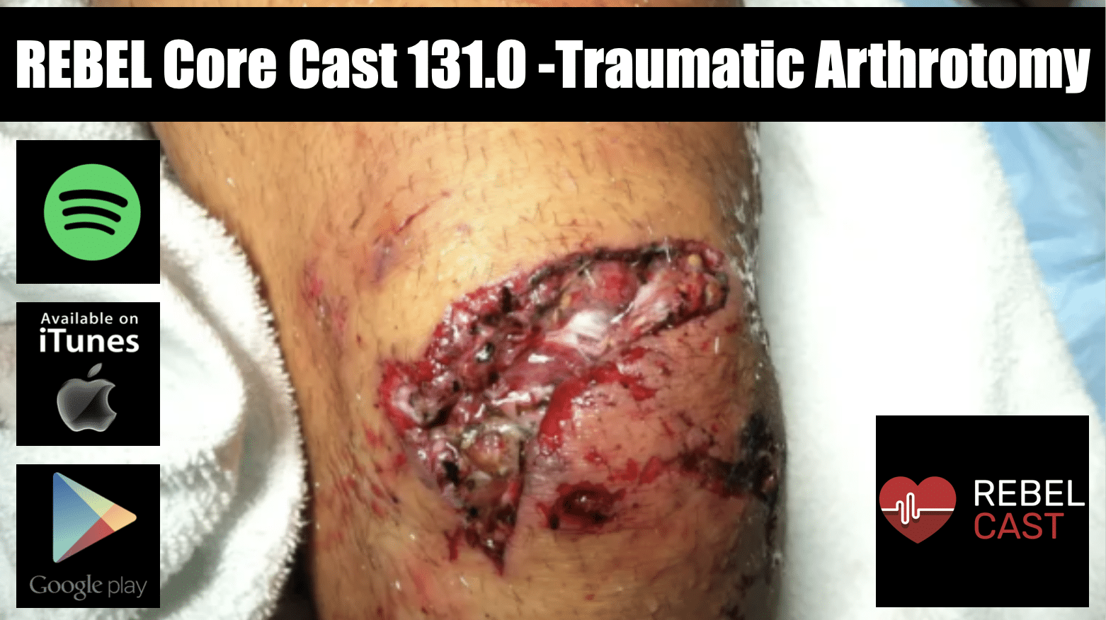
REBEL Core Cast 131.0 – Traumatic Arthrotomy
Manage episode 450809992 series 3381424

- Always suspect an open joint if there is a laceration, regardless of size, the lies over joint
- CT scan of the affected joint is widely considered to be the standard approach to evaluation but the saline load test may be useful in certain circumstances.
- Obtain emergency orthopedics consultation for all open joints and administer antibiotics and update tetanus in all patients
Definition: a deep laceration that extends into the joint capsule, exposing the intra-articular surface to the environment
- A laceration into the joint exposes the normally sterile intra-articular contents to external contamination
- Inoculation of the joint often results in septic arthritis
Physical Exam:
- Laceration over joint (can be variable in size)
- Local wound exploration may be sufficient in identifying the open joint
- Exam findings suspicious for joint capsule involvement:
-
- Air bubbles
- Extravasation of joint fluid – straw colored, viscous, sometimes oily in appearance
-
Diagnostic testing:
- Imaging:
- X-ray
- Limited ability to see air in joints but a reasonable first test
- CT scan
- Intra-articular air visualized on CT (Konda 2013)
- May be up to 100% sensitive for joint violation
- Study limited by small numbers, inclusion bias + inadequate gold standard
- May be considered the standard evaluation modality in many settings.
- Intra-articular air visualized on CT (Konda 2013)
- X-ray
- Saline load test
- Has mainly been supplanted by CT scan due to ease in obtaining, reported performance characteristics, consultant recommendation and difficulty in interpreting test.
- Useful if physical examination equivocal or plain radiographs non-diagnostic
- Technique (Video)
- Perform arthrocentesis of the joint with a large bore needle (18-20 gauge)
- Sterile saline is injected into the joint while passive movement is applied to the joint
- The laceration site is watched for saline extravasation indicating communication between the joint and external environment
-
- Sensitivity ranges from 34%-99% depending on the study, joint, and the amount of saline used to load the joint (Browning 2016)
- Methylene blue
- Aids in distinguishing a true positive from additional bleeding from the wound
- Recent studies suggest that the addition of methylene blue does not increase sensitivity if a sufficient amount of saline is used (Metzger 2012)
- Volume of fluid injected
- Varies depending on the joint in which you are injecting
- Higher volumes increase sensitivity but also increase pain for the patient
- Knee Joint (Keese 2007)
- 50 ml: Sensitivity of about 46%
- 194 ml: sensitivity of 95%
- Elbow Joint (Feathers 2011)
- 20 ml: Sensitivity of 86%
- 40 ml: Sensitivity of 95%
- Ankle Joint (Bariteau 2013)
- 7 ml: Sensitivity of 50%
- 30 ml: Sensitivity of 95%
ED Management:
- Reduce open fractures if present
- Irrigate grossly contaminated wounds in the ED
- Immobilize the joint to prevent further injury
- Obtain early orthopedic evaluation for joint exploration, and washout to be performed within 6-24 hours
- Tetanus prophylaxis
- Prophylactic antibiotics (best if given within 6 hours)
- Staph/strep coverage: 1st generation cephalosporin (i.e. cefazolin or cefuroxime)
- If risk factors for MRSA present, use agent with activity against MRSA (i.e. vancomycin)
- If significant soft tissue injury, add gram negative coverage like late generation cephalosporin, extended-spectrum penicillin, or aminoglycoside (i.e. gentamycin)
- If concern for fecal or clostridial infection, add high dose penicillin (i.e. zosyn)
- If seawater contamination and concern for vibrio vulnificus, add doxycycline
Post Peer Reviewed By: Salim R. Rezaie, MD (Twitter/X: @srrezaie)
The post REBEL Core Cast 131.0 – Traumatic Arthrotomy appeared first on REBEL EM - Emergency Medicine Blog.
29 afleveringen
Manage episode 450809992 series 3381424

- Always suspect an open joint if there is a laceration, regardless of size, the lies over joint
- CT scan of the affected joint is widely considered to be the standard approach to evaluation but the saline load test may be useful in certain circumstances.
- Obtain emergency orthopedics consultation for all open joints and administer antibiotics and update tetanus in all patients
Definition: a deep laceration that extends into the joint capsule, exposing the intra-articular surface to the environment
- A laceration into the joint exposes the normally sterile intra-articular contents to external contamination
- Inoculation of the joint often results in septic arthritis
Physical Exam:
- Laceration over joint (can be variable in size)
- Local wound exploration may be sufficient in identifying the open joint
- Exam findings suspicious for joint capsule involvement:
-
- Air bubbles
- Extravasation of joint fluid – straw colored, viscous, sometimes oily in appearance
-
Diagnostic testing:
- Imaging:
- X-ray
- Limited ability to see air in joints but a reasonable first test
- CT scan
- Intra-articular air visualized on CT (Konda 2013)
- May be up to 100% sensitive for joint violation
- Study limited by small numbers, inclusion bias + inadequate gold standard
- May be considered the standard evaluation modality in many settings.
- Intra-articular air visualized on CT (Konda 2013)
- X-ray
- Saline load test
- Has mainly been supplanted by CT scan due to ease in obtaining, reported performance characteristics, consultant recommendation and difficulty in interpreting test.
- Useful if physical examination equivocal or plain radiographs non-diagnostic
- Technique (Video)
- Perform arthrocentesis of the joint with a large bore needle (18-20 gauge)
- Sterile saline is injected into the joint while passive movement is applied to the joint
- The laceration site is watched for saline extravasation indicating communication between the joint and external environment
-
- Sensitivity ranges from 34%-99% depending on the study, joint, and the amount of saline used to load the joint (Browning 2016)
- Methylene blue
- Aids in distinguishing a true positive from additional bleeding from the wound
- Recent studies suggest that the addition of methylene blue does not increase sensitivity if a sufficient amount of saline is used (Metzger 2012)
- Volume of fluid injected
- Varies depending on the joint in which you are injecting
- Higher volumes increase sensitivity but also increase pain for the patient
- Knee Joint (Keese 2007)
- 50 ml: Sensitivity of about 46%
- 194 ml: sensitivity of 95%
- Elbow Joint (Feathers 2011)
- 20 ml: Sensitivity of 86%
- 40 ml: Sensitivity of 95%
- Ankle Joint (Bariteau 2013)
- 7 ml: Sensitivity of 50%
- 30 ml: Sensitivity of 95%
ED Management:
- Reduce open fractures if present
- Irrigate grossly contaminated wounds in the ED
- Immobilize the joint to prevent further injury
- Obtain early orthopedic evaluation for joint exploration, and washout to be performed within 6-24 hours
- Tetanus prophylaxis
- Prophylactic antibiotics (best if given within 6 hours)
- Staph/strep coverage: 1st generation cephalosporin (i.e. cefazolin or cefuroxime)
- If risk factors for MRSA present, use agent with activity against MRSA (i.e. vancomycin)
- If significant soft tissue injury, add gram negative coverage like late generation cephalosporin, extended-spectrum penicillin, or aminoglycoside (i.e. gentamycin)
- If concern for fecal or clostridial infection, add high dose penicillin (i.e. zosyn)
- If seawater contamination and concern for vibrio vulnificus, add doxycycline
Post Peer Reviewed By: Salim R. Rezaie, MD (Twitter/X: @srrezaie)
The post REBEL Core Cast 131.0 – Traumatic Arthrotomy appeared first on REBEL EM - Emergency Medicine Blog.
29 afleveringen
Alle afleveringen
×Welkom op Player FM!
Player FM scant het web op podcasts van hoge kwaliteit waarvan u nu kunt genieten. Het is de beste podcast-app en werkt op Android, iPhone en internet. Aanmelden om abonnementen op verschillende apparaten te synchroniseren.


